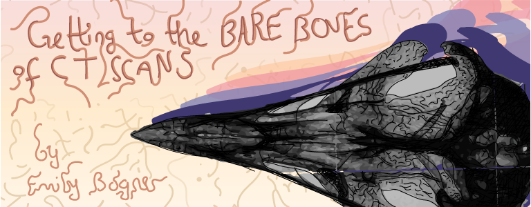
Toolbox: Getting to the Bare Bones of CT Scans
Computerized tomography (CT) scanners may be most notable in the medical field as tools to diagnose muscle and bone disorders, guide procedures, or detect and monitor diseases; but CT scanners serve a myriad of other purposes both in and outside of the biological sciences. A major benefit to this type of imaging is its non-invasive nature, with the ability to construct three-dimensional images of internal structures without destruction.
The capabilities of a CT scanner may sound very similar to that of a typical X-ray. However, there is one fundamental difference between the two: whereas X-rays send radiation in only one direction to produce a single two-dimensional image, CT scanners provide X-ray measurements from multiple angles, which are then compiled to create a three-dimensional image. You can think of the object being scanned like a loaf of bread composed of many thin slices stacked next to each other. A planar X-ray shows only one slice of the bread, but a CT scanner recreates the entire loaf. The comprehensive images produced by CT scanners can greatly accelerate scientific exploration of a particular structure, without having to take it apart.
Assistant professor Dr. Jack Tseng from the Department of Integrative Biology (IB) brought a new Nanotom CT scanner to Berkeley’s campus in August 2020, and it is already contributing to some groundbreaking research. Second-year IB PhD student Jennifer Hoeflich is utilizing this technology to study ear bones in fish. Fish are particularly good models for studying sound production, which can give researchers insights into hearing loss and deafness in humans. The Nanotom system is the first industrial-strength, high-resolution CT system on the Berkeley campus capable of producing high quality images of structures that are as small as 0.6 microns—shorter than the length of a bacterium! The Nanotom is perfect for studying tiny structures almost invisible to the naked eye.
To be scanned, each fish skull is placed on a rotating stage inside the CT scanner, where a special digital X-ray detector is located directly opposite the X-ray source. When Hoeflich scans a fish, the transmitter sends out X-rays that pass through the skull and are picked up by the detectors on the other side. Depending on the amount of X-ray that is absorbed in the skull’s scales, cartilage, soft tissue, and bone, different amounts of X-ray pass through the fish and reach the detectors. This process allows the CT scanner to determine varying material densities and other structural properties in a single scan, allowing for more structural data to be collected than with a planar X-ray. The stage the fish skull sits on then rotates 360 degrees so the X-ray can compile an image from every angle. Once a scan is complete, the density information is transmitted to a computer where, depending on the size of the object, hundreds or thousands of slices (remember our loaf of bread?) will be compiled into a single three-dimensional image.
Hoeflichs’ research only scratches the surface of what is possible with the Nanotom. Dr. Tseng’s hope for the Nanotom is to involve and benefit any researcher needing to capture fine, three-dimensional geometric details of a variety of biological and non-biological specimens. He hopes to work across campus departments to link quantitative information and analyses across multiple biological scales, from centimeters to nanometers. The IB department is just one slice of the campus’ intellectual community—Tseng wants his research program to draw on all of Berkeley’s interests and expertise to accelerate research across the sciences.
-------
Emily Bogner is a graduate student in integrative biology.
Design by Emily Carvalho
This article is part of the Fall 2021 issue.